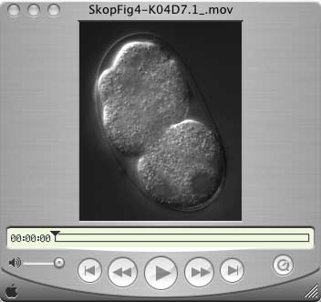
Caption: RACK1
RNAi-treated embryo. This movie shows cell division without the presence of
a key protein. The nucleus is the dark circle inside the cell; as the life
cycle goes on, the nucleus replicates and each of the two nuclei move to opposite
ends of the cell. The outer membrane furrows and seems to pinch in, but ultimately
does not close itself off to form two complete cells. Instead, the cell is
left with two complete nuclei, which is a fatal flaw.
Video by: courtesy Ahna Skop
Date: May 2004
QuickTime video

Caption:
RACK1 RNAi-treated embryo. This movie shows cell division without the presence
of a key protein. The nucleus is the dark circle inside the cell; as the life
cycle goes on, the nucleus replicates and each of the two nuclei move to opposite
ends of the cell. The outer membrane furrows and seems to pinch in, but ultimately
does not close itself off to form two complete cells. Instead, the cell is
left with two complete nuclei, which is a fatal flaw.
Video by: courtesy Ahna Skop
Date: May 2004
QuickTime video

Caption: CHO
cells dividing with isolated midbodies surrounding them. The cells and midbodies
are stained with anti-actin (red), anti-tubulin (green) and DAPI (blue). This
image shows Chinese hamster ovary cells in the last stages of division. The
red outer membrane is complete around each new cell, while the green midbody
still remains between them. Isolated midbodies are also pictured in green around
the cells to show the organelles in more detail.
Photo by: courtesy Ahna Skop
Date: May 2004
High-resolution 300 DPI JPEG

Caption: CHO
cells dividing. The cells are stained with anti-actin (red), anti-tubulin (green)
and DAPI (blue). This image shows two Chinese hamster ovary cells in the last
stages of division. The red outer membrane is complete around each new cell,
while the green midbody still remains between them.
Photo by: courtesy Ahna Skop
Date: May 2004
High-resolution 300 DPI JPEG

